
Postoperative Wound Dehiscence in a Dog with Adjunctive Laser Therapy
CASE SUMMARY
| Patient | Huahua |
| Species | Canine (Pomeranian) |
| Age | 3 years |
| Clinical presentation | Postoperative wound dehiscence with marginal skin necrosis |
| Treatment | 17 sessions of adjunctive LYRA laser therapy alongside conventional wound care |
| Laser system | AILUCS LYRA Five-Wavelength Veterinary Laser |
| Final recorded appearance | Most of the wound surface appeared epithelialized and covered with crust |
Case Overview
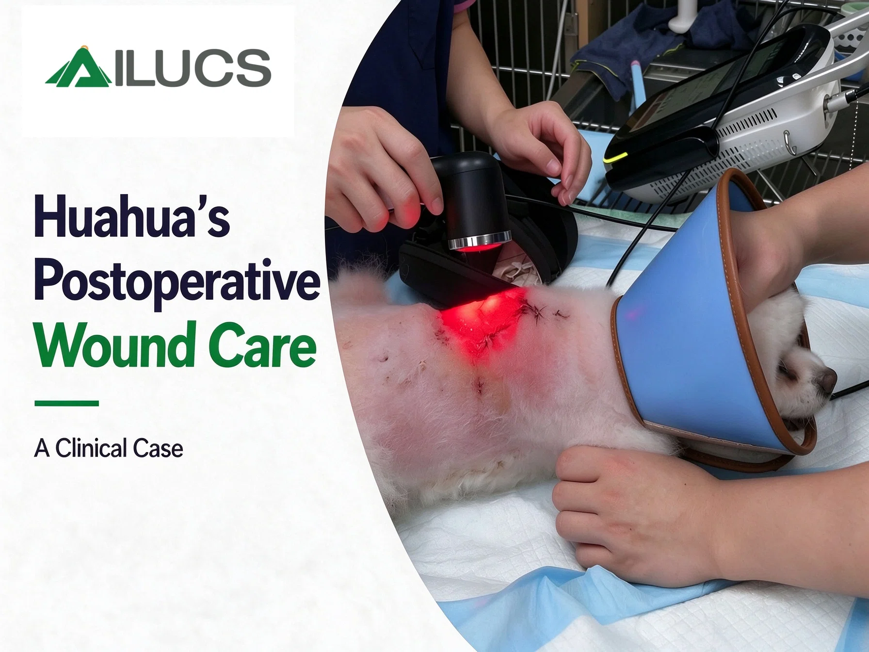
Huahua, a three-year-old Pomeranian, was presented with postoperative wound dehiscence at a previous surgical site. Necrotic tissue was visible along the wound margins, and the wound remained open. Adjunctive LYRA laser therapy was incorporated into the care plan alongside conventional wound care. This case documents the recorded laser protocol and changes in wound appearance across 17 sessions from September 27 to November 4, 2025.
Initial Wound Assessment
At the initial assessment, dark necrotic crust and exposed suture material were visible along the wound margins, with partial separation of the wound edges. The wound bed was red and moist, with yellow-white fibrinous exudate at parts of the wound base. Purple discoloration and mild swelling were visible in the surrounding skin.
Huahua wore an Elizabethan collar to prevent licking and chewing at the wound.
The wound-care plan focused on managing necrotic tissue and exudate, supporting granulation tissue formation, and monitoring wound-edge epithelialization and contraction.For a broader understanding of how veterinary laser therapy may be incorporated into wound management, explore our guide to veterinary laser therapy for pet wound healing, including the biological processes involved in tissue repair and the role of photobiomodulation as an adjunct to conventional wound care.
Postoperative Laser Therapy Protocol
Device: LYRA Five-Wavelength Veterinary Laser System
Clinical indication: Postoperative open wound with marginal skin necrosis
Treatment area: Open wound and surrounding tissue
Treatment course: 17 sessions across three phases
| Phase | Sessions | Wavelengths | Mode |
|
Phase 1 Sep 27–29, 2025 |
3 | 650 + 810 + 915 nm | Pulsed |
|
Phase 2 Oct 2–12, 2025 |
6 | 650 + 810 + 915 + 980 nm | Pulsed |
|
Phase 3 Oct 13–Nov 4, 2025 |
8 | 810 + 915 + 980 + 1064 nm | Pulsed |
Wavelength selection rationale: The wavelength combination was adjusted in stages so that treatment followed the changing clinical priorities of each healing phase. In Phase 1, when necrotic tissue, fibrinous exudate and active surface inflammation predominated, the 650 + 810 + 915 nm combination directed red and near-infrared (NIR) coverage to the superficial wound bed and wound margins. In Phase 2, as the wound bed became more evenly granulating, 980 nm was added to extend coverage into the deeper wound bed and periwound tissue during the proliferative phase. In Phase 3, as epithelialization advanced and the open area shrank, the protocol replaced the superficial 650 nm with 1064 nm, the longest wavelength and deepest-penetrating among those applied, to sustain repair support in the deeper tissue while the surface became largely covered by epithelium and crust.
Clinical note: These settings document the protocol used in this individual case and should not be interpreted as a standardized treatment prescription. Treatment parameters should be selected by a qualified veterinarian according to the patient, target tissue, and clinical objectives.
Laser therapy was applied to the open wound and surrounding tissue alongside conventional wound care. No laser session was performed on October 17, 2025; wound dressing was performed on that day.
Photographic Wound Progression
Baseline | Before the First Recorded Laser Session
The baseline photographs show an open wound with necrotic crust and exposed suture material along the wound margins. The wound bed was red and moist, with yellow-white fibrinous exudate at the wound base. Purple discoloration and mild swelling were visible in the surrounding skin.
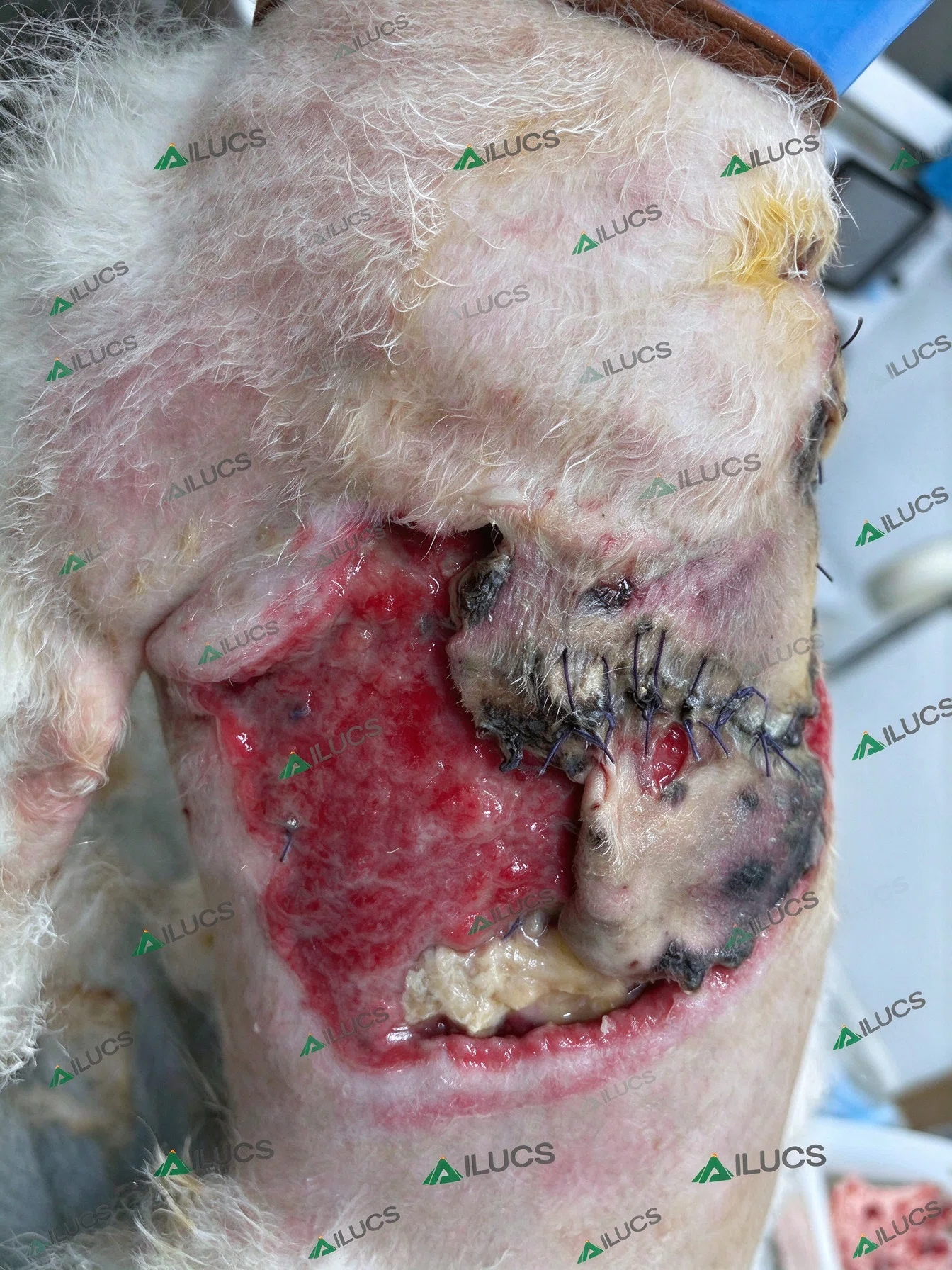
Figure 1. Baseline appearance before the first recorded laser session.
October 2, 2025 | After Three Recorded Laser Sessions
By October 2, 2025, the wound bed appeared bright red, with granulation tissue distributed more evenly across the surface. Exudate had decreased, and the wound margins appeared more defined. The wound remained open at this stage.
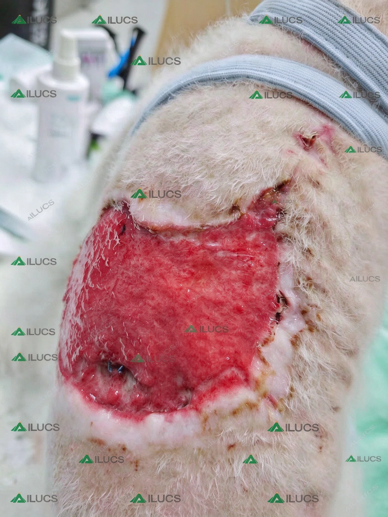
Figure 2. Granulation tissue visible across the wound bed on October 2, 2025.
October 13, 2025 | Wound Appearance After Phase 2
By October 13, the wound surface remained red and glossy, with markedly less exudate than at the initial assessment. Epithelialization was visible along the wound margins, extending toward the center, while the granulation tissue appeared more compact and smooth. The central area had not yet fully closed.
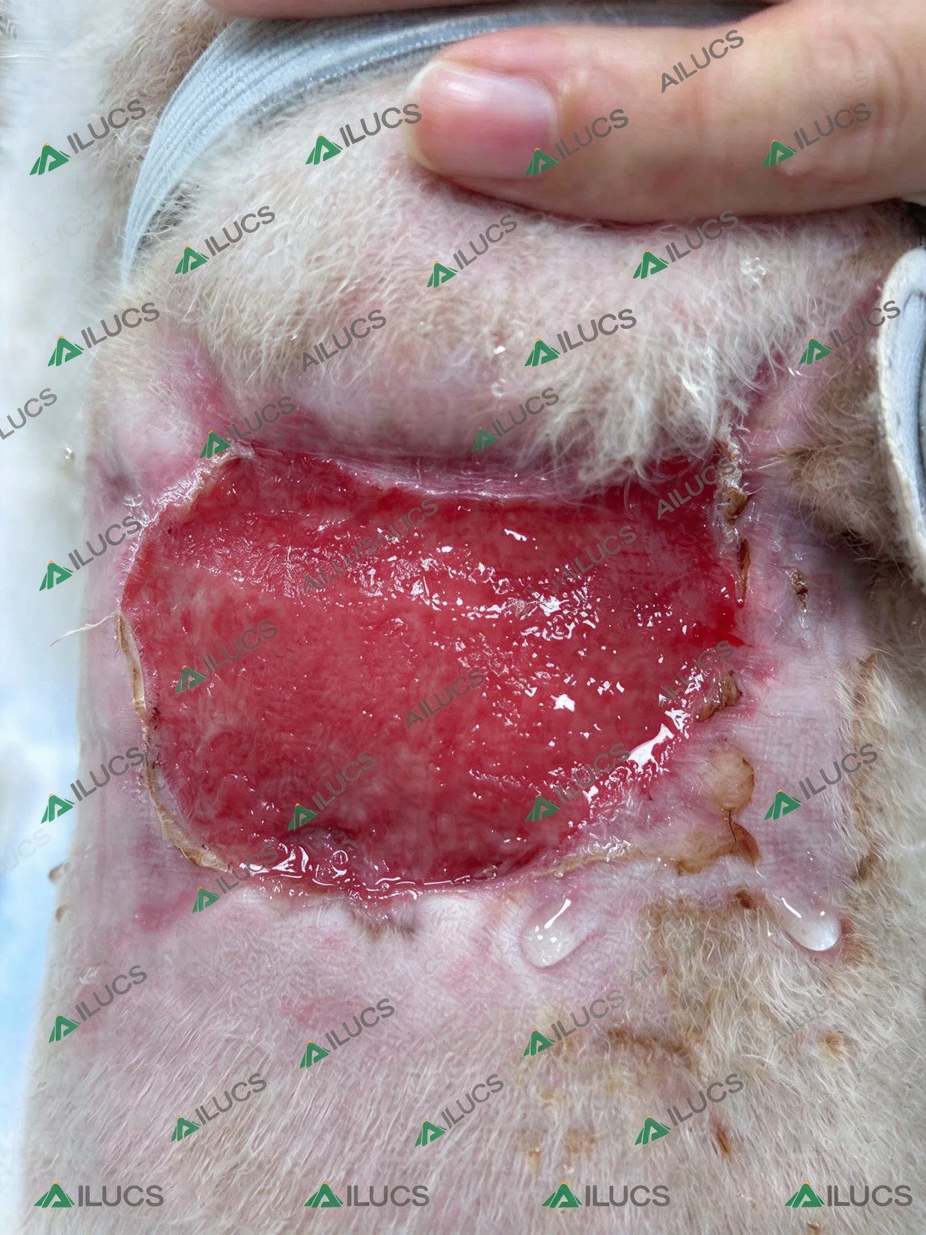
Figure 3. Epithelialization visible along the wound margins on October 13, 2025.
October 29–November 4, 2025 | Later Wound Progression
In the later photographs, the wound continued to decrease in size and the epithelial margin became more defined. On October 29, the wound was substantially smaller, although the central area remained open. The final photograph dated November 4 shows that most of the wound surface appeared epithelialized and covered with crust. Exact session counts for individual photographs in this late interval are not specified in the available photographic record.
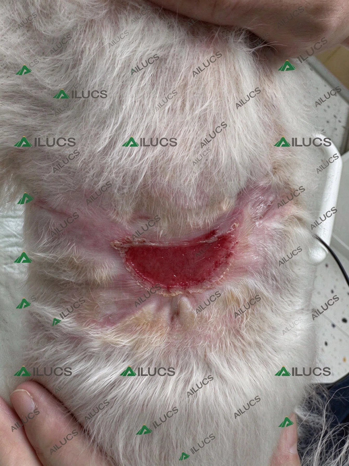
Figure 4. Wound appearance on October 29, 2025, with a smaller wound surface and a more defined epithelial margin.
Final Recorded Outcome
At the final recorded visit on November 4, 2025, most of the wound surface appeared epithelialized and covered with crust.
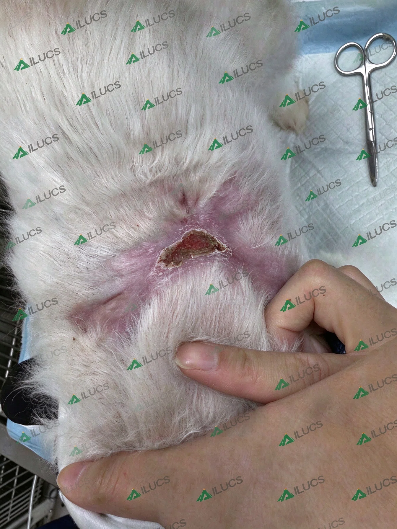
Figure 5. Final recorded wound appearance on November 4, 2025.
Clinical Interpretation
The serial photographs document changes in Huahua's postoperative wound, from necrotic wound margins and fibrinous exudate to a more evenly granulating surface, visible epithelialization, and a smaller wound area. LYRA laser therapy was incorporated into the wound-care plan alongside conventional wound care throughout the documented treatment period.
For comparison, another canine clinical case documents the use of adjunctive LYRA laser therapy following postoperative incision redness and swelling. While Huahua's case involved wound dehiscence and marginal skin necrosis, this case records a closed surgical incision with postoperative inflammation. Explore our related clinical case on canine postoperative incision laser therapy.
Recommendations
For clinical practices managing similar postoperative wound dehiscence cases, this case illustrates how a single multi-wavelength platform — the AILUCS LYRA Five-Wavelength Veterinary Laser — can support phase-appropriate laser therapy: wavelength combinations were adjusted across the healing course without changing devices, from superficial red and NIR coverage in the acute stage to the addition of deeper-penetrating wavelengths (980 nm, then 1064 nm) during granulation and epithelialization.
When using the LYRA system for wound management, clinicians are advised to record the clinical indication, healing phase, wavelength combination, session frequency and serial photographs for objective review, and to select treatment parameters case by case under the direction of a qualified veterinarian.
Veterinary Disclaimer: This case is presented for professional education and product-use context. It does not establish a universal treatment protocol or guarantee the same outcome in other patients. Diagnosis, wound management, medications, and laser parameters should be determined by qualified veterinary professionals.