
Adjunctive Laser Therapy for Dorsal Skin Necrosis and Wound Myiasis in a Stray Dog
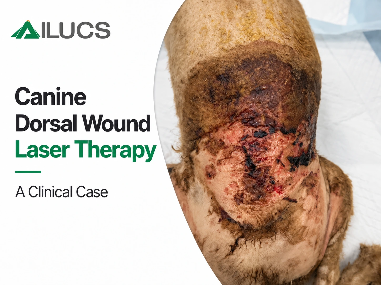
Case Overview
Patient Details
Species: Canine
Age: Unknown
Sex: Male
Body weight: 9 kg
Presenting Complaint
A rescued stray dog was presented with a malodorous dorsal wound and maggot infestation. Clipping revealed extensive skin damage, localized necrosis, and an open wound with exudation and signs of inflammation. Management centered on debridement, infection management, and ongoing wound care, with photobiomodulation therapy (PBMT) added as an adjunct.
Clinical Examination
The dog was in fair general condition but had poor body condition. Examination revealed an extensive area of dorsal skin injury that was more clearly defined after clipping.
The wound showed the following features:
Superficial necrotic tissue, including localized gray-black areas.
Erythema and swelling at the wound margins, with disruption of skin integrity.
A moist wound surface with exudate and scant blood-tinged discharge.
Local signs suggestive of infection, with associated tissue breakdown.
Numerous maggots within the wound after removal of the overlying hair, consistent with wound myiasis.
Epidermal loss and exposed superficial tissue in some areas.
Initial Wound Assessment
No extensive muscle exposure was evident on initial inspection. Subsequent observations focused on necrotic tissue, exudate, wound margins, and coverage of the wound surface.
For a broader overview of wound assessment and the role of supportive treatment, see our guide to veterinary laser therapy for pet wound healing
Pretreatment Assessment
Given the necrosis, tissue loss, contamination, and myiasis, the case was managed as a complex cutaneous wound. Priorities were to remove maggots and devitalized tissue, manage infection, protect the wound, and support subsequent repair. PBMT was used as an adjunct to this approach.
Treatment Goals
The goals were to remove maggots and devitalized tissue, manage infection, reduce local discomfort, protect the wound, and support progressive wound closure. Subsequent treatment and care were to be adjusted according to wound appearance and the dog's tolerance.
Treatment Protocol
Wound Preparation
Before PBMT, the affected area was clipped, maggots and contaminants were removed, and the wound was cleaned and debrided. These measures were combined with infection management.
PBMT was then incorporated into ongoing wound care. Protective dressings and continued wound care were provided after treatment.
For additional guidance on treatment timing and application considerations, see when laser therapy may be appropriate after a pet injury
Photobiomodulation Therapy
Device: LYRA Veterinary Laser System
Wavelength combination: 650, 810, 915, 980, and 1064 nm
The treatment sequence consisted of one application of the skin wound protocol followed by one application of the muscle preset for adjunctive periwound treatment.
To understand the biological principles behind this adjunctive approach, read our introduction to photobiomodulation in veterinary medicine
Skin Wound Protocol
Treatment area
Skin wounds over the back and rump.
Parameters
| Parameter | Setting |
| Preset indication | Skin wound |
| Preset wound size | Large (60–150 cm²) |
| Preset stage | Acute |
| Output power | 1.8 W |
| Average power | 0.9 W |
| Mode | Pulsed |
| Protocol duration | 5 min 48 s (348 s) |
| Total energy per protocol | 313.2 J |
Application technique
The entire wound area was treated using a scanning technique, with the applicator held approximately 1–2 cm above the wound. The applicator was moved evenly over the area without prolonged stationary exposure at any one site.
Muscle Preset for Adjunctive Periwound Treatment
Treatment area
Periwound areas over the back and rump.
Parameters
| Parameter | Setting |
| Preset tissue | Muscle |
| Species | Canine |
| Preset treatment site | Back |
| Preset weight category | 5–10 kg |
| Preset stage | Acute |
| Output power | 6.7 W |
| Average power | 3.3 W |
| Mode | Pulsed |
| Protocol duration | 3 min 56 s (236 s) |
| Total energy per protocol | 790.6 J |
Note: Duration refers to the complete run of each protocol. Output power, average power, and total energy are reported as displayed on the device.
Application technique
Treatment was directed at the periwound area.
A contact technique was used.
The applicator was positioned perpendicular to the treatment area.
The applicator was moved slowly over the treatment region to cover the surrounding tissue.
Direct pressure on the open wound was avoided.
Treatment Progress
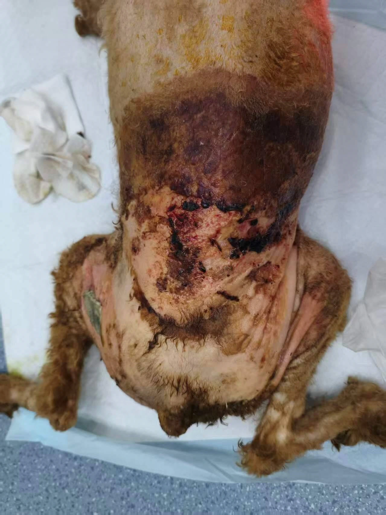
Day 0 Before Adjunctive Laser Therapy
At the start of care, the wound showed necrosis, contamination, and maggot infestation. PBMT was added to ongoing wound management after initial wound treatment.
Day 1 After the First Treatment
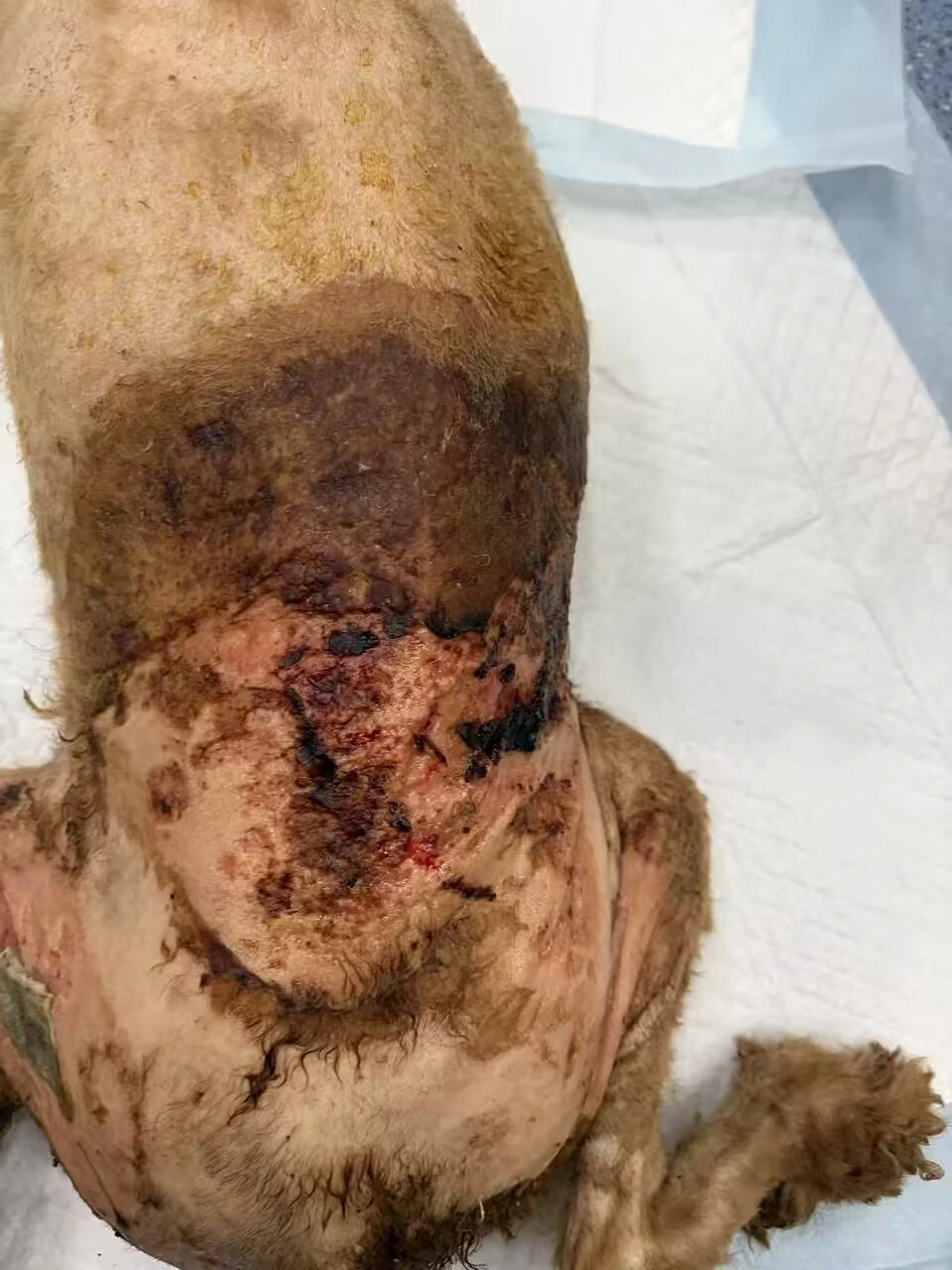
After the first treatment, dark necrotic areas and local tissue injury remained visible. Adjunctive PBMT and wound care continued.
Day 2 After the Second Treatment
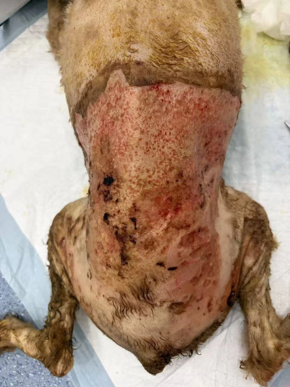
Extensive skin injury remained over the back. The wound appeared red, with areas of crusting and exudation.
Day 21 Interim Wound Record
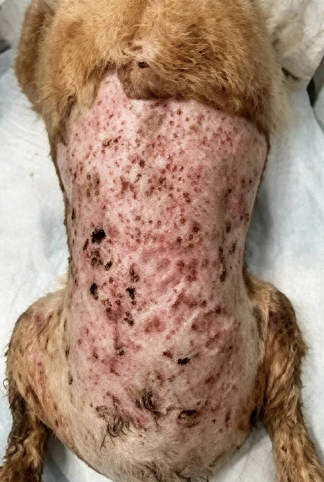
Compared with the early images, the wound showed an improved overall appearance and greater coverage of the affected surface. Localized erythema and scattered crusts remained visible.
Adjustment of Treatment Frequency
In the early phase, necrosis, exudation, and inflammation were more pronounced, and PBMT was generally performed two to three times per week. After Day 21, as the wound stabilized and local signs improved, the frequency was reduced to one to two treatments per week. Routine wound care continued.
Day 42 Final Wound Photograph
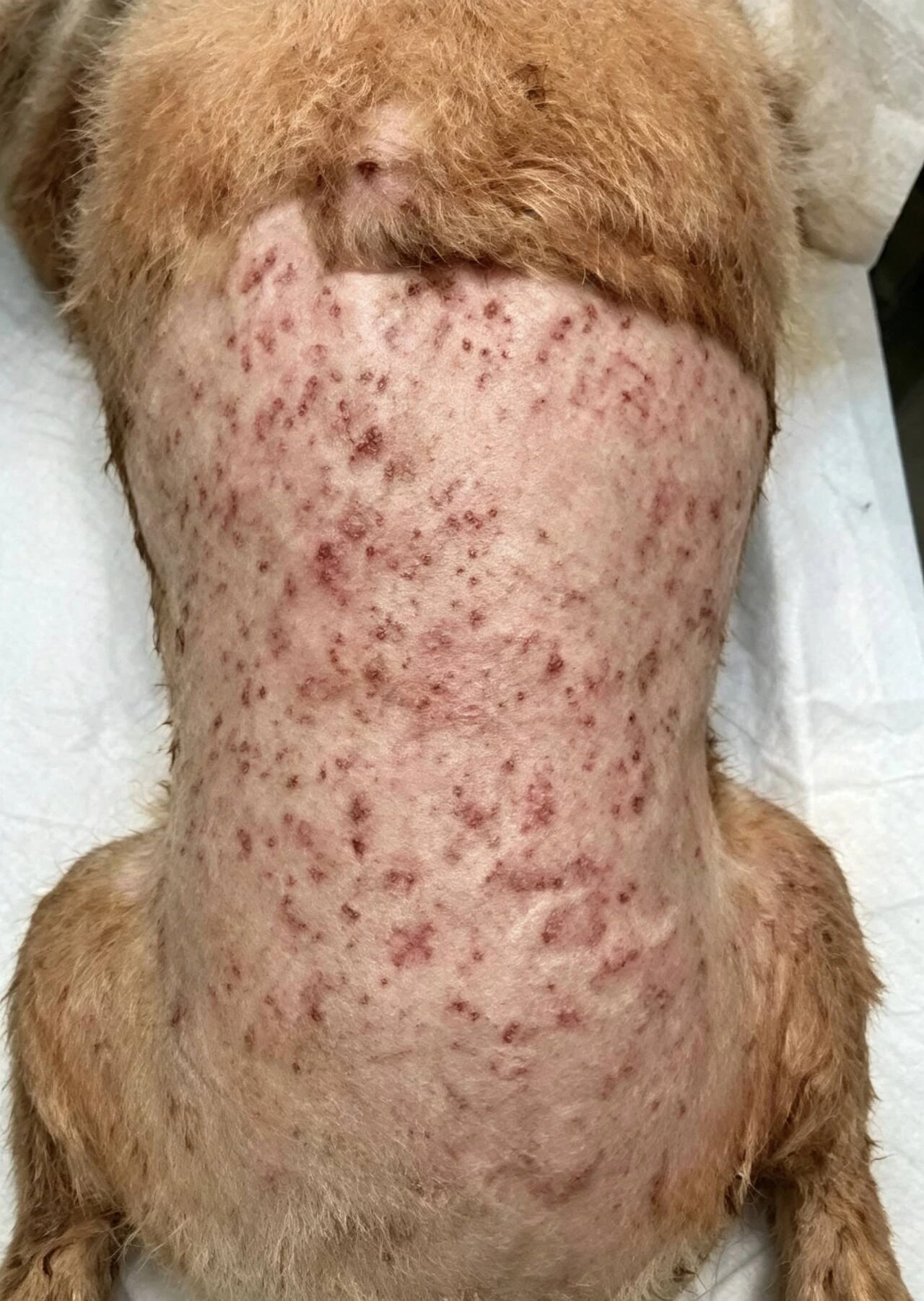
The final photograph showed marked improvement from baseline. Most of the affected surface appeared covered, and early hair regrowth was visible in some areas. Scattered erythema and minor crusting remained.
Observed Outcome
During combined management with debridement, infection management, protective dressings, routine wound care, and adjunctive PBMT, wound appearance improved over time. Serial records showed less necrotic tissue and exudate than in the early phase, increased coverage of the affected surface, and early hair regrowth in some areas.
The dog remained cooperative during the documented treatment period, and no obvious adverse effects were observed.
Discussion and Limitations
This case illustrates the use of PBMT as an adjunct to comprehensive wound management. Improvement occurred alongside debridement, infection management, protective dressings, and routine care. The contribution of each intervention cannot be separated, and this case alone cannot establish whether laser therapy shortened the healing period.
This single case was documented mainly through serial photographs and clinical descriptions. The observations reflect only this dog's progress during the recorded period. They cannot quantify the independent effect of laser therapy or establish a treatment protocol for general use.
For another documented wound-care case involving adjunctive LYRA laser therapy, explore our feline wound healing clinical case.
Clinical Disclaimer
This report is intended for professional education and case discussion. The parameters are specific to this case and are not a universal prescription. This information does not replace veterinary diagnosis, debridement, infection management, or individualized treatment assessment.