
Canine Postoperative Incision Laser Therapy: A Clinical Case

Case Summary for Publication
This clinical case documents a dog that developed marked redness and swelling around a closed surgical incision after a routine sterilization procedure. The report follows the visible appearance of the incision across three adjunctive LYRA Veterinary Laser sessions and an owner-provided follow-up two days after the dog returned home. Its value lies in the longitudinal record: a defined treatment protocol, serial photographs, and a short-term follow-up image that allows the surgical site to be compared over time. Because the available record does not include standardized wound scores, laboratory measurements, or an untreated comparison, the case should be read as a descriptive clinical observation rather than proof that laser therapy alone produced the observed recovery.
1. Case Overview
Species: Dog
Treatment site: Surgical incision following a routine spay/neuter procedure
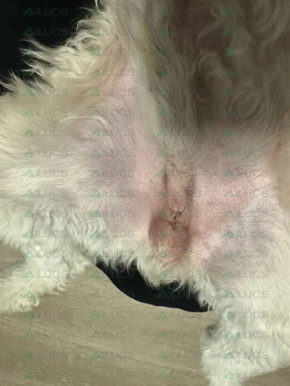
Following sterilization surgery, the dog developed marked redness and swelling around the incision, with a localized inflammatory response. The incision remained closed, and sutures were still visible.
Adjunctive treatment was provided using the LYRA Veterinary Laser, for a total of three laser therapy sessions.
2. Pre-Treatment Assessment
Before laser therapy, pronounced redness and swelling were visible around the surgical incision. The surrounding skin showed a clear erythematous change, together with localized tissue swelling. Based on the local clinical appearance, the main postoperative concern was redness, swelling, and a localized inflammatory response around the incision.
Treatment goals:
Reduce local redness, swelling, and inflammatory response.
Support postoperative tissue repair.
Promote recovery of the surgical incision.
3. Treatment Protocol
Device: LYRA 5 Veterinary Laser System
Clinical indication: Postoperative incision redness and swelling
Treatment area: Surgical incision and surrounding tissue
Number of treatments: 3 sessions
| Parameter | Setting |
| Wavelengths | 650 nm + 810 nm + 915 nm + 980 nm + 1064 nm |
| Power | 1.6 W |
| Average power | 0.8 W |
| Mode | Pulsed |
| Treatment time | 1 min 36 sec |
| Total energy | 76.8 J |
| Treatment head | 30 mm spot-size treatment head |
Case-specific protocol note. These settings document the protocol used in this individual case and should not be interpreted as a universal treatment prescription. Treatment parameters should be selected by the responsible veterinary professional according to the patient, tissue target, device, and clinical goals.
Laser therapy was applied to the surgical incision and surrounding tissue based on the local condition, with serial observation of tissue changes throughout the treatment period.
4. Treatment Progress
Before Treatment
Marked redness and localized tissue swelling were visible around the surgical incision.
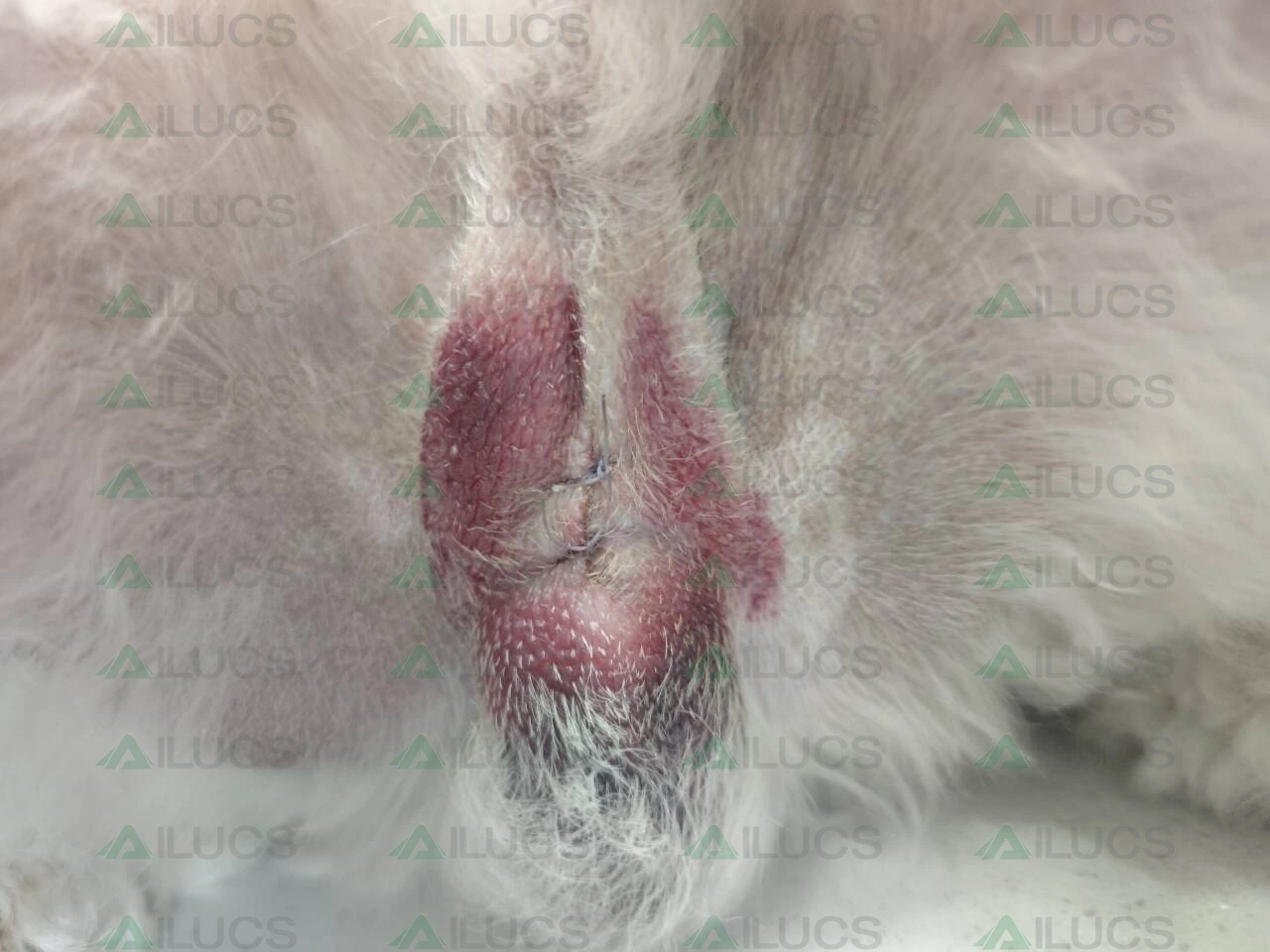
After the First Treatment
After the first laser therapy session, marked redness was still visible around the surgical incision.
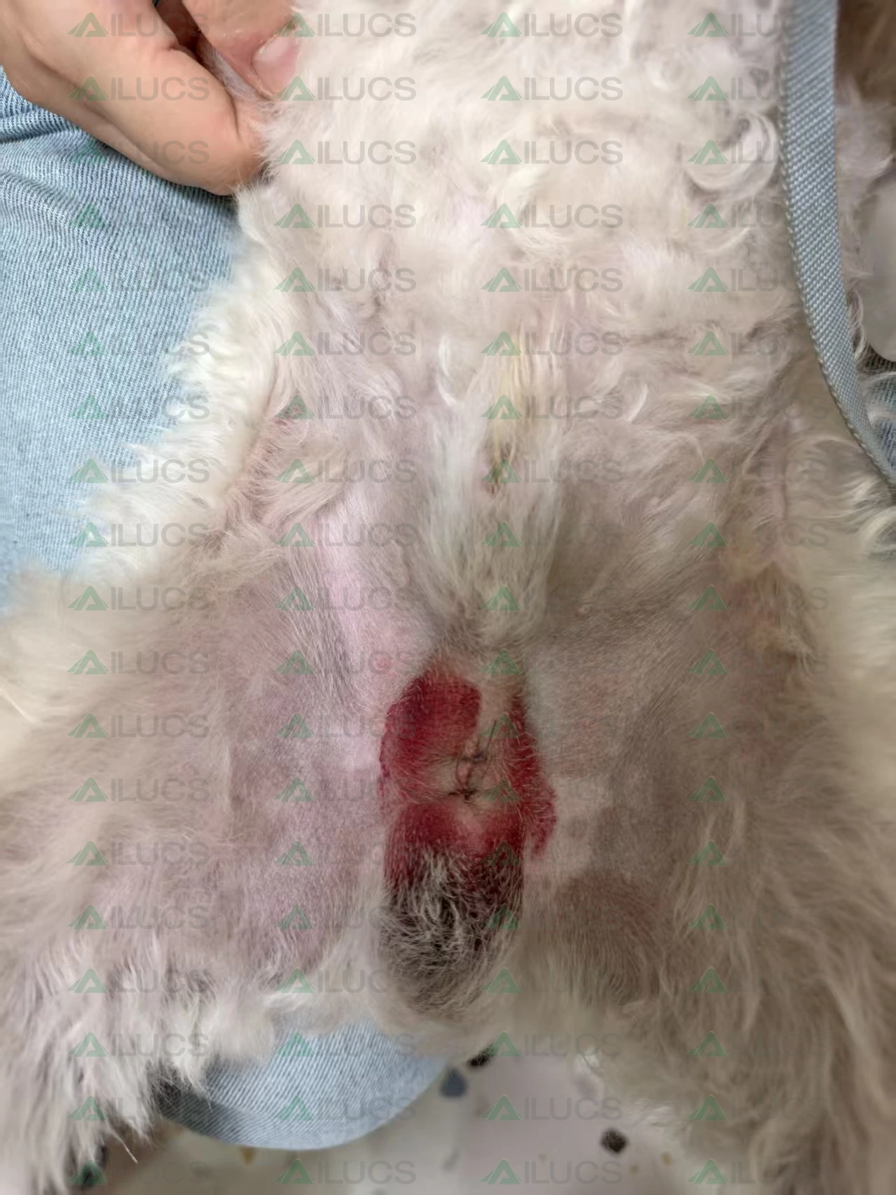
After the Second Treatment
After the second laser therapy session, the incision remained closed, although localized redness was still present.
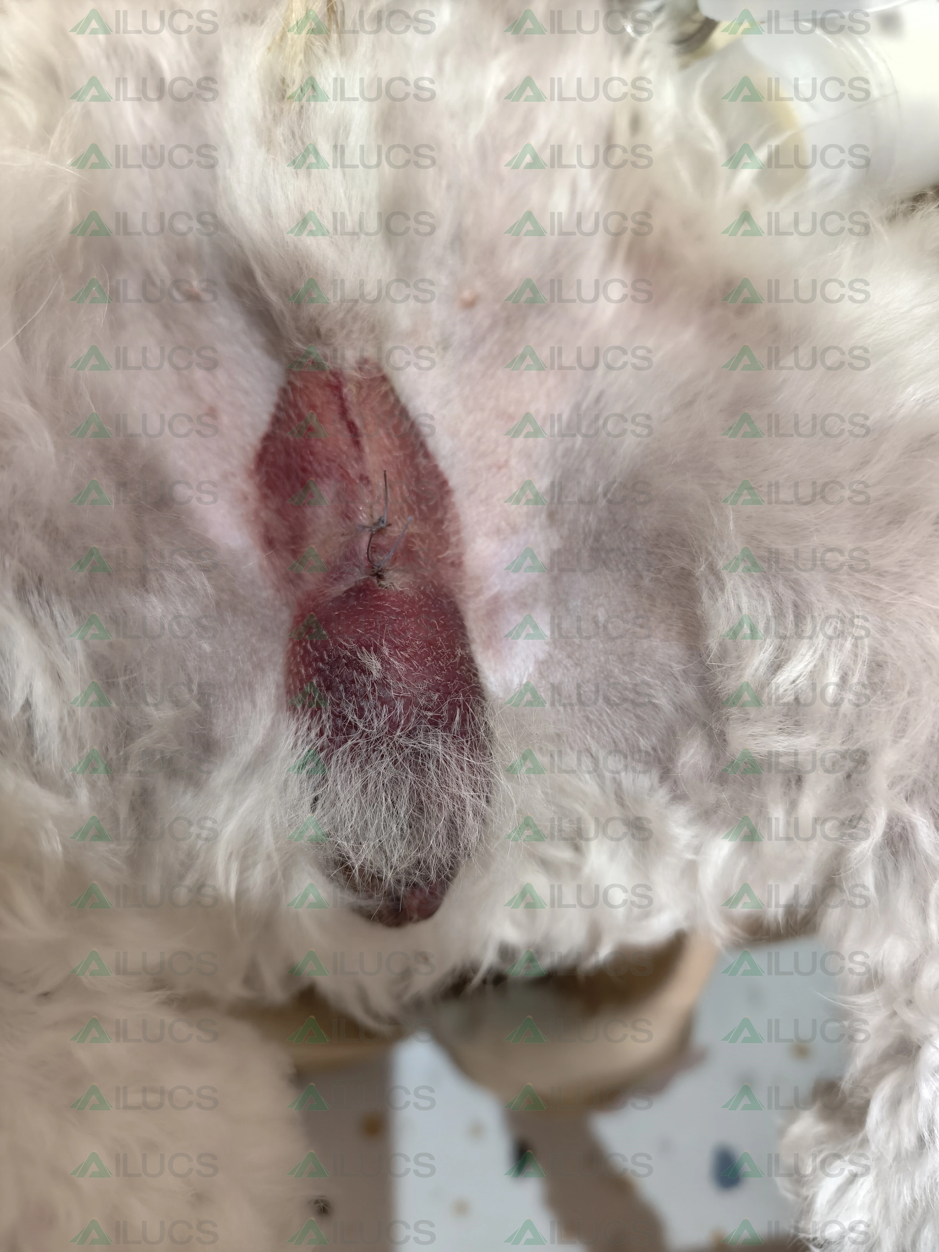
After the Third Treatment
After the third laser therapy session, the overall condition of the treated area remained stable.
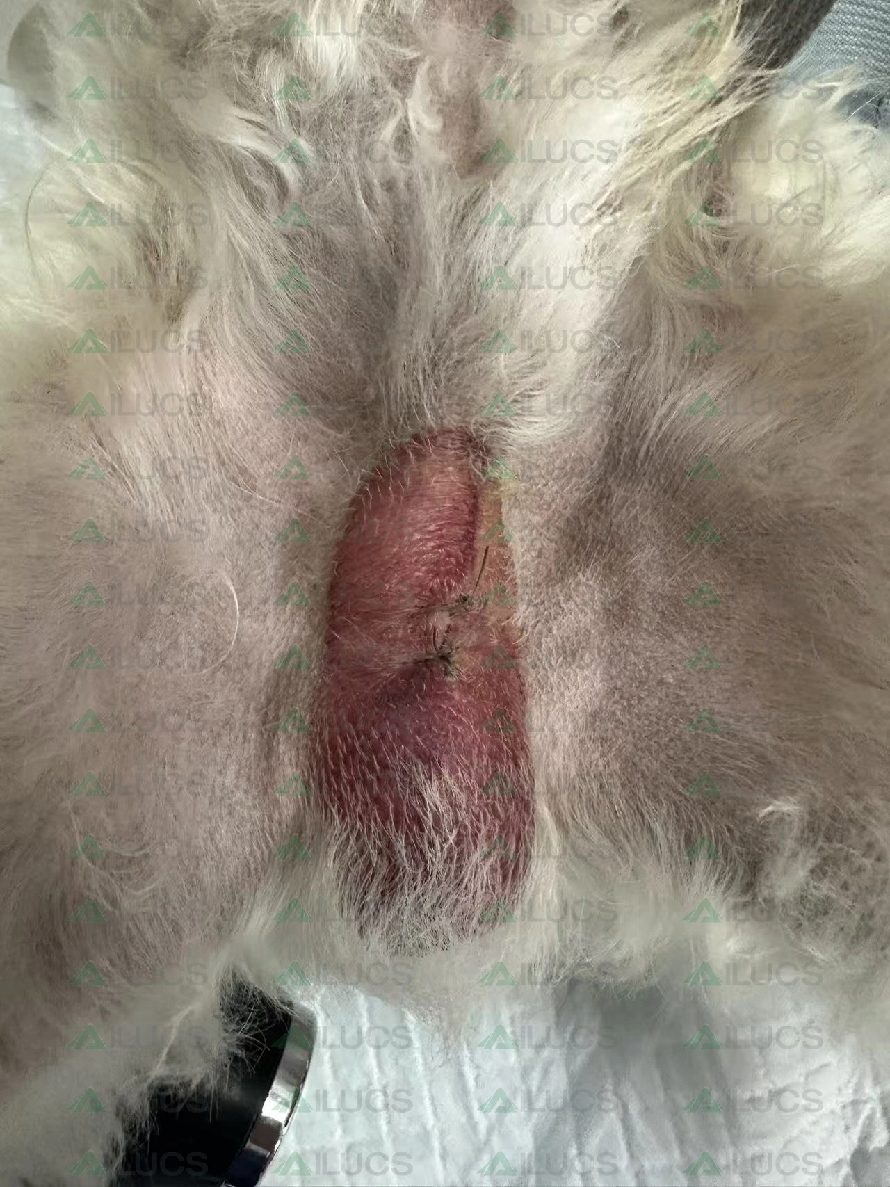
Two-Day Follow-Up
Two days after the dog returned home, the owner provided follow-up photographs. The pronounced redness seen before treatment had largely subsided, the local skin color had visibly improved, and the surgical area appeared to be recovering well.
5. Clinical Observation Summary
Before treatment, the main clinical findings were marked redness and swelling around the postoperative incision, together with a localized tissue reaction.
The dog received three sessions of adjunctive laser therapy. Changes in local redness and swelling were monitored throughout the treatment course, and treatment was continued with ongoing observation of the surgical area.
At the two-day follow-up after the third treatment, owner-provided photographs showed a clear improvement in the appearance of the surgical area.
Overall observations:
The surgical incision remained closed throughout the observation period.
No obvious wound dehiscence was observed during treatment.
After three laser therapy sessions, the treated area showed progressive recovery.
At the two-day follow-up, the previously marked redness had substantially decreased.
The overall appearance of the surgical area had improved substantially.
6. Clinical Interpretation
Across the serial photographs, the incision remained closed during the observed treatment period. Redness was still visible after the early sessions, while the owner-provided two-day follow-up showed a visibly improved surgical-site appearance compared with the pre-treatment image. These observations should be interpreted within the context of normal postoperative recovery and the dog’s broader surgical aftercare. The source record does not include a standardized wound-scoring system, laboratory data, or a control condition, so it cannot determine how much of the change was attributable specifically to laser therapy. In this case, laser therapy was used as an adjunctive component of postoperative management while the local tissue response was monitored over time.
7. Clinical Takeaway
The strongest feature of this case is its visual and parameter-documented timeline: a clearly described postoperative concern, three recorded laser sessions, and a short-term owner follow-up. Presented this way, the case offers a practical example of how veterinary laser therapy may be incorporated into postoperative care without implying that the same protocol or outcome will apply to every patient.
Veterinary disclaimer: This case is provided for professional education and product-use context. It does not establish a universal treatment protocol or guarantee the same outcome in other patients. Diagnosis, postoperative care, wound management, and laser parameters should be determined by qualified veterinary professionals.