
FHO Recovery in Dogs and Cats
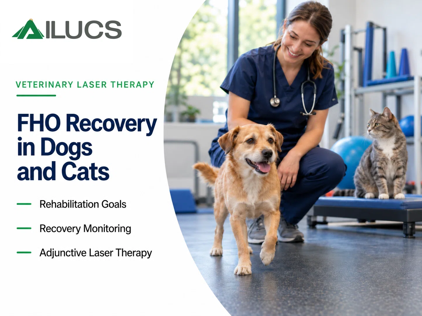
Recovery after femoral head ostectomy (FHO) involves more than healing the incision. As the tissues around the hip adapt, the dog or cat must regain comfortable use of the operated leg. Standing independently, bearing weight and returning to daily activities are separate milestones, each offering useful information about progress.
This guide explains what FHO changes at the hip, how rehabilitation progresses and what owners can monitor at home. It also discusses the role of adjunctive laser therapy and links the postoperative pathway to our canine hip dysplasia guide and Paipai’s feline clinical case.
How FHO changes the hip
FHO usually involves removal of the femoral head and neck. The surrounding soft tissues support a functional false joint, eliminating painful contact between the affected bone surfaces without restoring a normal ball-and-socket joint. Some patients retain changes in hip motion, limb length or gait. Rehabilitation is recommended to preserve movement and support useful function.
The incision may heal before the patient has regained comfortable, consistent use of the leg. Follow-up therefore needs to assess both wound healing and hip function.
The link with canine hip dysplasia
FHO is one surgical option for selected dogs with hip dysplasia, but it is not an automatic next step when conservative management falls short. An orthopedic assessment should determine which procedure, if any, is appropriate for the individual patient.
For non-surgical management, read our companion guide to laser therapy for hip dysplasia in dogs and non-surgical management. Once FHO has been performed, the focus shifts to postoperative limb use and adaptation to the altered hip.
How long does FHO recovery take
Recovery progresses over weeks, with the pace and extent of improvement varying between patients. A dog or cat may stand independently before walking comfortably or tolerating its usual activity. The surgeon’s examination and the patient’s response should guide progression, rather than a fixed calendar.
ACVS notes that patients with FHO as their only injury may typically be allowed more activity after the first two weeks than those recovering from some other hip procedures. This comparison does not authorize unrestricted exercise. Additional injuries and the surgeon’s instructions remain central to the activity plan.
Ask the clinic when the incision will be checked and when limb function will be reviewed. At each appointment, confirm which activities are now allowed, which restrictions remain and what changes warrant an earlier visit. Independent standing alone is not clearance for running or jumping.
Planning rehabilitation before discharge
Before going home, obtain written instructions for medication, incision protection, permitted activity and follow-up. Ask the veterinary team to demonstrate any exercises or handling techniques you will need to use. If a support aid is recommended, learn how to fit it without placing pressure on the surgical area.
The plan should explain both how to protect the patient and how to encourage prescribed movement. FHO, fracture fixation and total hip replacement require different approaches to recovery. Confirm the procedure performed and any additional injuries before starting home exercises.
Rebuilding use of the operated leg
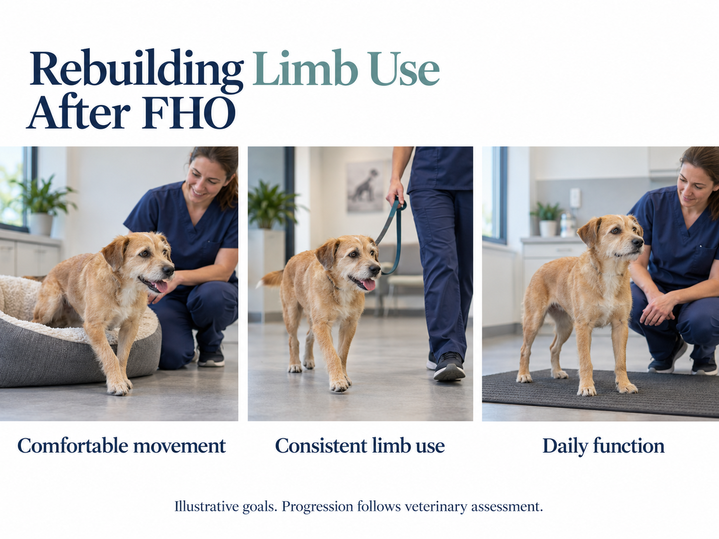
After FHO, rehabilitation addresses movement at the altered hip and the muscles that support it. The stages below describe functional goals, not a timetable or exercise prescription. Progression should follow veterinary assessment.
| Stage | FHO rehabilitation goal | What the team reviews |
| Comfortable supported movement | Protect the surgical area while starting the approved return to limb use | Pain during position changes and the assistance needed to rise |
| Consistent use of the operated leg | Progress from occasional paw placement toward tolerated weight bearing | Use of the leg during approved movement and comfortable hip motion |
| Strength around the altered hip | Develop muscle support and control for the functional false joint | Muscle condition, gait and tolerance before increasing exercise |
| Sustainable daily function | Use the limb comfortably within the patient’s realistic goals | Remaining gait or motion limitations and clearance for particular activities |
At follow-up, the rehabilitation clinician can assess hip motion, muscle condition and use of the operated leg. At home, perform only exercises the team has demonstrated. Forcing hip movement or increasing activity to make the patient use the leg may worsen discomfort and make the cause of stalled progress harder to identify.
Adapting the home environment
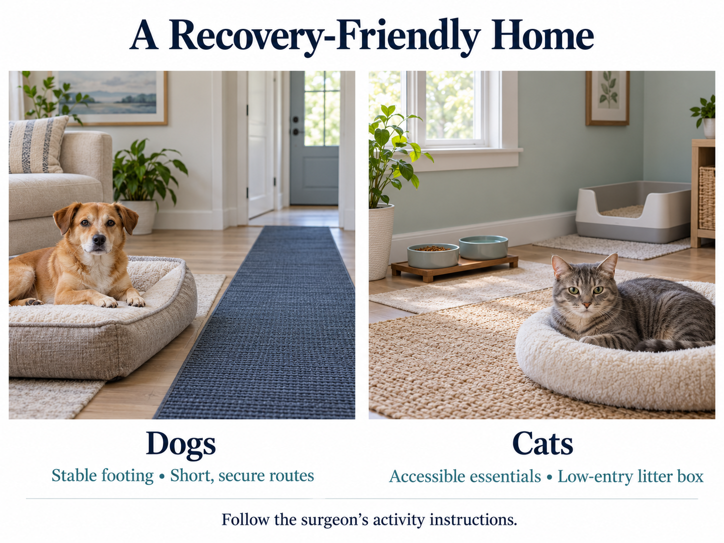
For dogs, plan a short, secure route between the resting area and the toileting area. Discuss stairs and any need for a support aid with the clinic. Provide stable footing and prevent sudden bursts of activity through doorways or toward other pets. The duration and pace of leash walks should follow the discharge instructions.
For cats, keep food, water, a resting place and an accessible litter box on the same level. Ask whether a lower litter-box entrance would help. Limit access to furniture as directed by the surgeon, and provide a quiet hiding place where the cat can still be monitored. These adjustments help maintain access to essential resources during recovery.
Observe movement within the approved routine rather than repeatedly testing the hip. For a cat, note whether entering the litter box or changing resting position becomes easier. For a dog, record how much assistance is needed for toileting. Use the same permitted activities to compare progress over time.
The role of laser therapy after FHO
A veterinarian may consider laser therapy when postoperative discomfort limits a prescribed rehabilitation activity. The treatment objective should be specific, such as improving tolerance of assisted rising or approved walking. AAHA includes photobiomodulation among rehabilitation modalities, but its guidance does not establish a specific recovery benefit after FHO.
Review changes in limb use alongside adjustments to medication and exercise. Improved comfort is useful to document, although it does not show which component of treatment produced the change. Laser therapy should complement the postoperative plan. Treatment timing, device settings and eye protection should follow the veterinarian’s assessment and the device instructions.
Paipai’s postoperative rehabilitation
Paipai, a five-month-old cat with a right femoral head fracture, underwent surgical removal of the fractured femoral head. The published case describes seven inpatient laser therapy sessions with LYRA as part of postoperative management. Independent standing was observed after the third session.
This was an early sign of functional progress, not evidence of complete hip recovery or proof that laser therapy caused the improvement. It also does not establish a treatment schedule for other cats. Because the published account does not confirm the full operative terminology, the procedure is described here using the documented information.
Read Paipai’s postoperative laser rehabilitation case for the photographs, treatment records and case timeline.
Monitoring function and recognizing setbacks
An incision review and a functional assessment answer different questions. A closed incision does not establish restored hip motion, strength or comfortable weight bearing. Similarly, briefly placing the paw on the floor is different from consistently using the leg during an approved walk.
Ask the team to assess how the operated leg contributes to standing and walking, whether hip movement is comfortable and whether muscle condition is improving. These findings provide a fuller picture of recovery than incision photographs alone.
At home, agree with the clinic on an approved activity to monitor, such as rising from a resting position or reaching the toileting area. Record the assistance needed, use of the operated leg and any discomfort afterward. Short videos may help at follow-up if requested; do not add stairs, jumping or longer walks to test recovery. Contact the veterinarian promptly if pain increases, the incision opens or develops discharge, or the patient stops using a leg it had begun to use. Persistent avoidance of the operated leg calls for reassessment, rather than simply adding exercise or laser sessions.
The goal is comfortable use of the operated leg within the patient’s realistic functional limits. The surgeon, rehabilitation clinician and owner should agree on the next permitted activity and review the patient’s response before progressing.
Veterinary disclaimer
This article provides general educational information. Diagnosis, surgery, medication, rehabilitation and laser parameters must be determined by qualified veterinary professionals for each patient. Clinical case observations do not establish treatment efficacy or guarantee similar outcomes.
References
[1] American College of Veterinary Surgeons. Femoral Capital Physeal Fractures. Source
[2] American College of Veterinary Surgeons. Canine Hip Dysplasia. Source
[3] AAHA. 2022 Pain Management Guidelines. Nonpharmacologic Modalities for Pain Management. Source
[4] AILUCS. Paipai’s Postoperative Laser Rehabilitation. A Clinical Case. Published October 9, 2026. Source