
Paipai's Postoperative Laser Rehabilitation: A Clinical Case
 |
CASE SNAPSHOT |
|
| Patient | Paipai | |
| Age | 5 months | |
| Diagnosis | Right femoral head fracture | |
| Surgery | Surgical excision of the fractured femoral head (exact operative term to be confirmed from hospital record) |
|
| Rehabilitation | Seven inpatient laser therapy sessions as an adjunct to postoperative care |
|
| Early functional milestone | Independent standing documented after 3 sessions | |
1. Case Overview
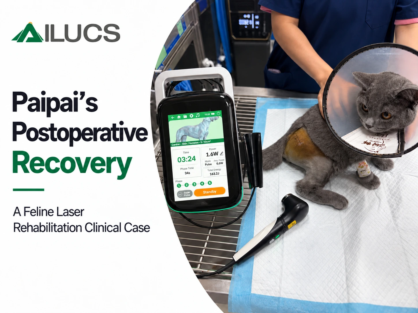
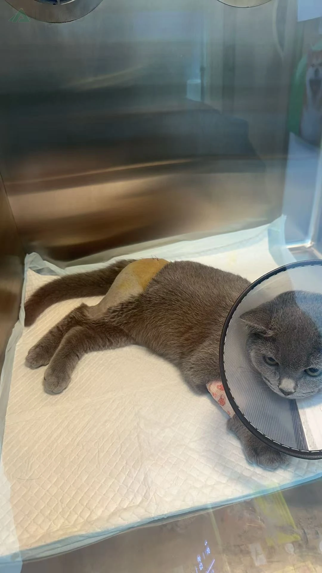
Paipai is a 5-month-old cat diagnosed with a fracture involving the right femoral head. According to the case information provided, the fractured femoral head was surgically excised. After surgery, Paipai remained hospitalized for postoperative care and rehabilitation. Veterinary laser therapy was incorporated as an adjunctive modality during this period, with seven treatment sessions documented through photographs and video.
Veterinary laser therapy was incorporated as an adjunct to postoperative care, with recovery documented through serial incision photographs, treatment footage, and functional observations.
2. Initial Diagnosis and Preoperative Imaging
The preoperative radiograph below was used to document the orthopedic injury before surgery. Patient- and hospital-identifying text has been removed from the publishing copy; the radiographic anatomy itself has not been intentionally altered.
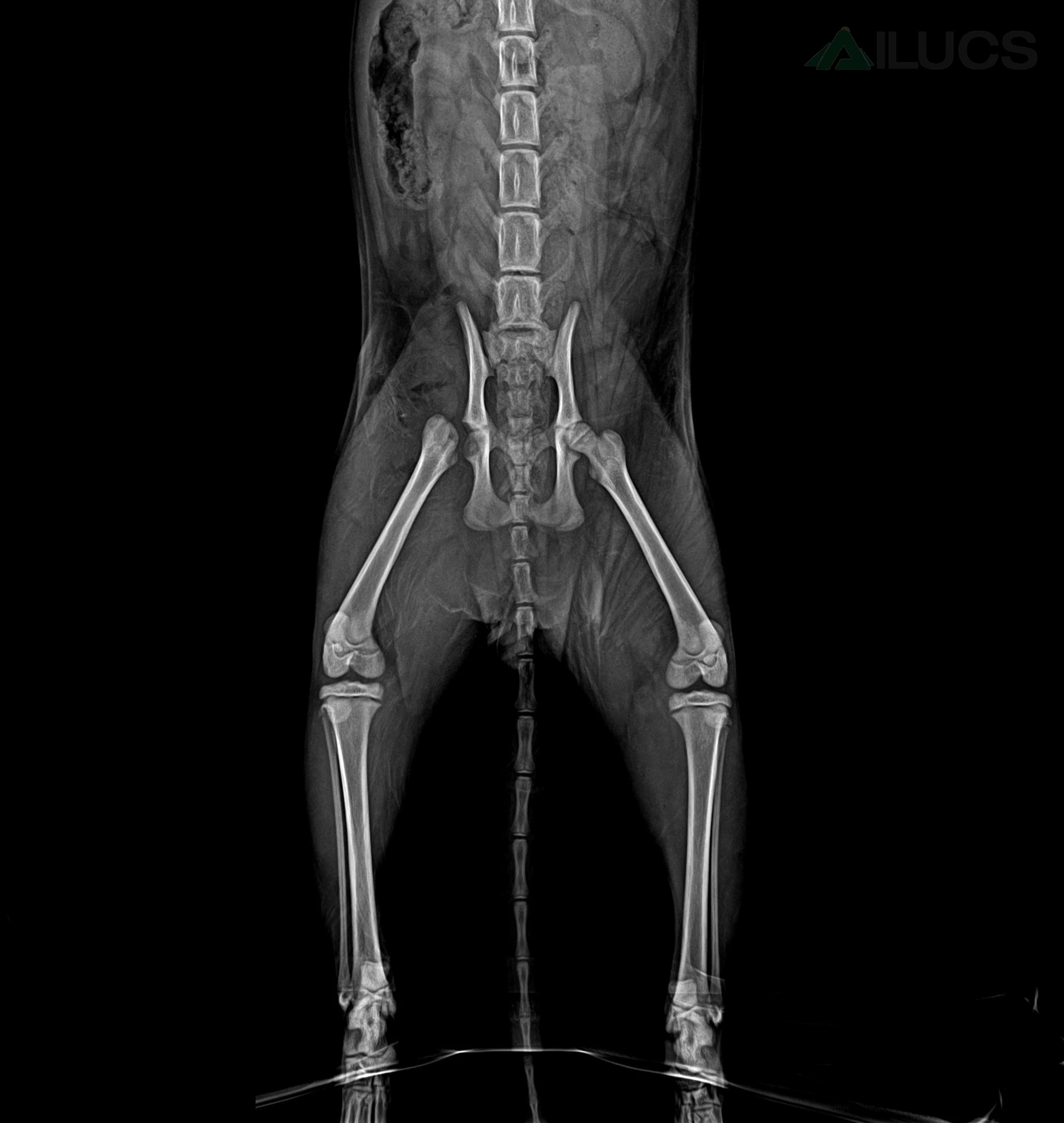
Figure 1. Preoperative radiograph of Paipai showing the hip region. The documented injury involved the right femoral head.
3. Postoperative Laser Therapy Protocol
The first three-session recovery video provides the following case-specific settings for the LYRA Five-Wavelength Veterinary Laser:
| Target area | Mode | Power | Average power | Total energ |
| Skin / incision | Pulsed | 1.6 W | 0.8 W | 163.2 J |
| Bone / hip region | Pulsed | 8.8 W | 4.4 W | 404.8 J |
Clinical Note: The laser settings documented in this case were specific to Paipai's postoperative rehabilitation and should not be considered a standardized treatment protocol. Treatment parameters must be determined by a qualified veterinarian based on the patient's condition, target tissue, and clinical objectives.
4. Early Rehabilitation: Sessions 1-3
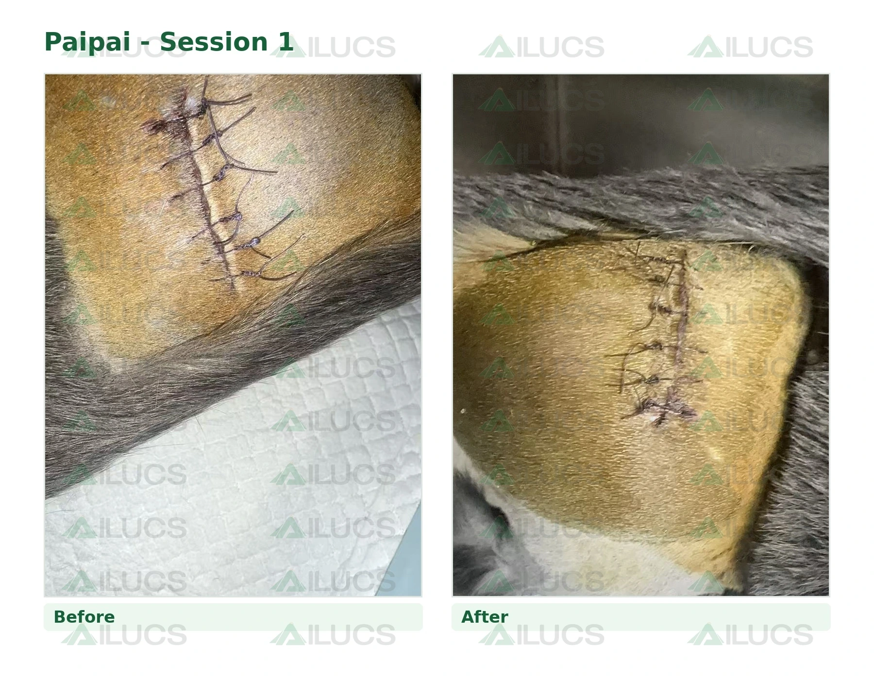
The video record documents the first three postoperative treatment sessions. Rather than interpreting immediate pre-/post-session photographs as proof of tissue healing, the case is best understood as a longitudinal recovery sequence across the hospitalization period.
Session 1: Postoperative baseline incision appearance was documented. The incision/skin region and hip region were treated according to the case protocol.
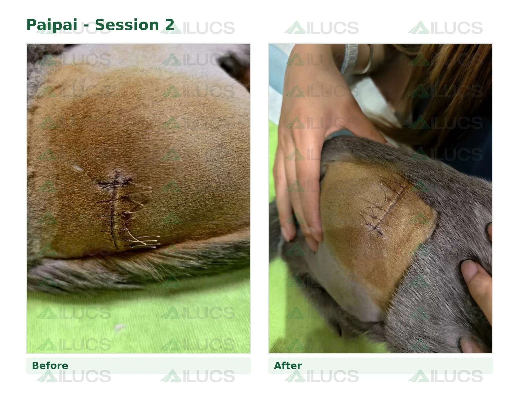
Session 2: The video describes the incision edges as healing with improving soft-tissue appearance, while Paipai was observed rising more easily with less guarding.
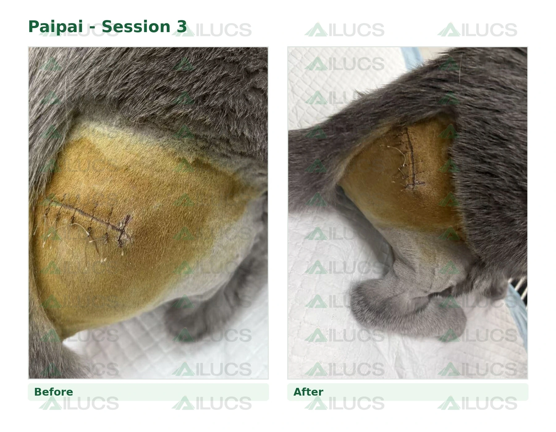
Session 3: The video describes the incision as closed well with improved surrounding tissue and documents more independent rising. Independent standing is shown in the early-recovery video after the third session.
Watch Paipai’s early recovery video (Sessions 1-3):https://youtube.com/shorts/Fcb96us94pc?si=cMaYJJ1rV9HY_ko7
5. Continued Inpatient Rehabilitation: Sessions 4-7
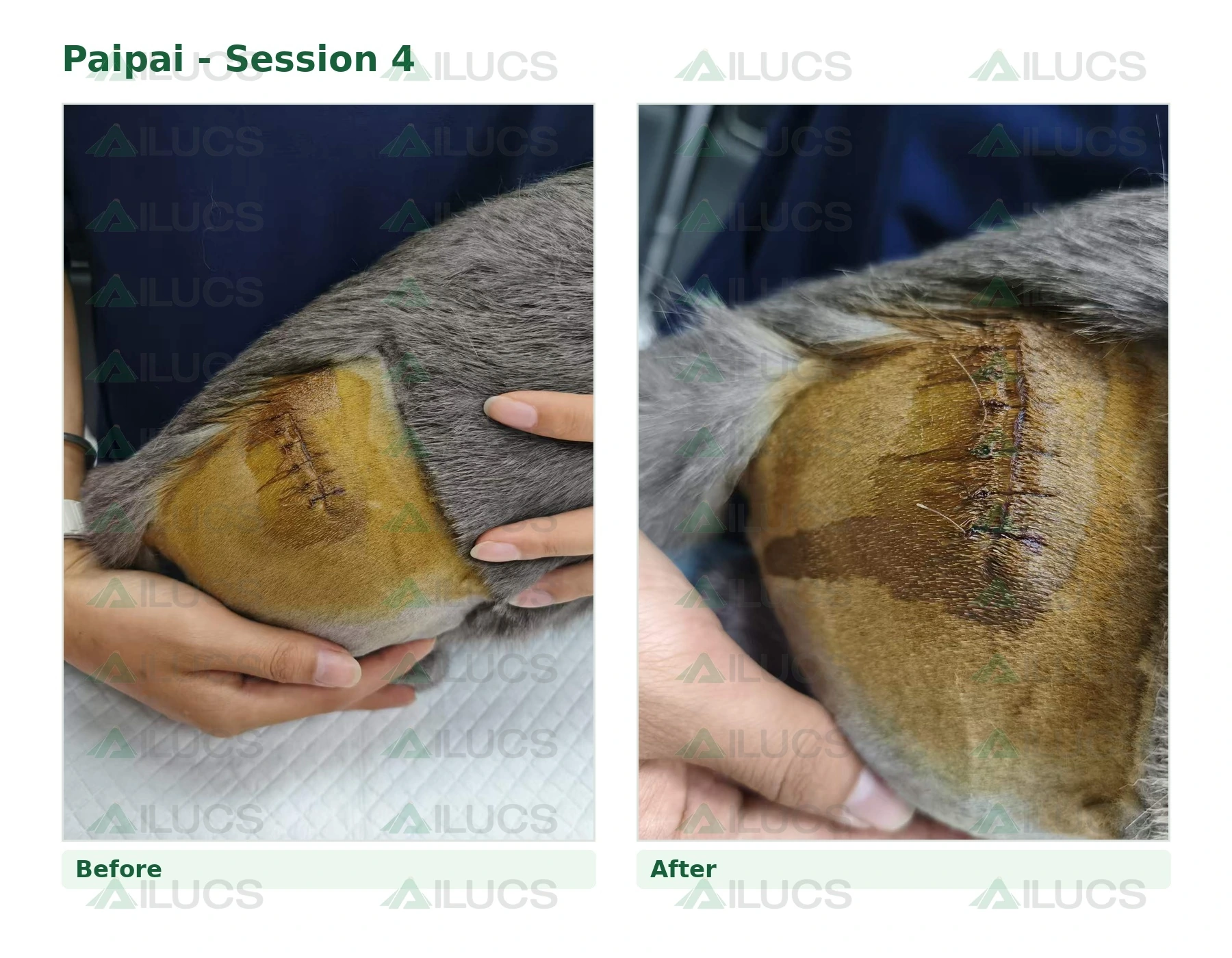
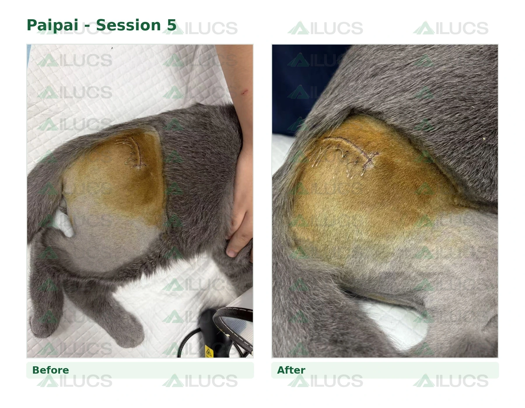
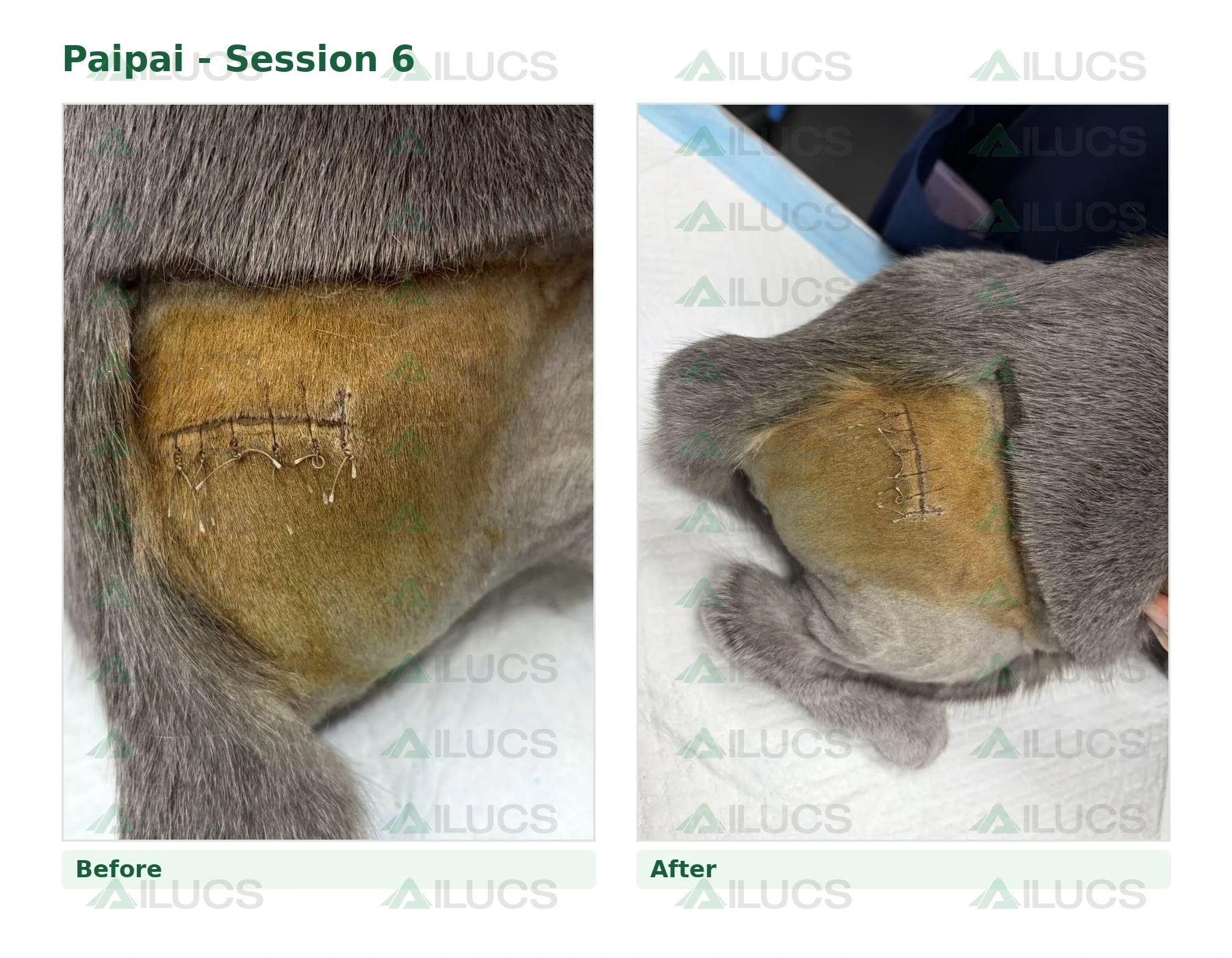
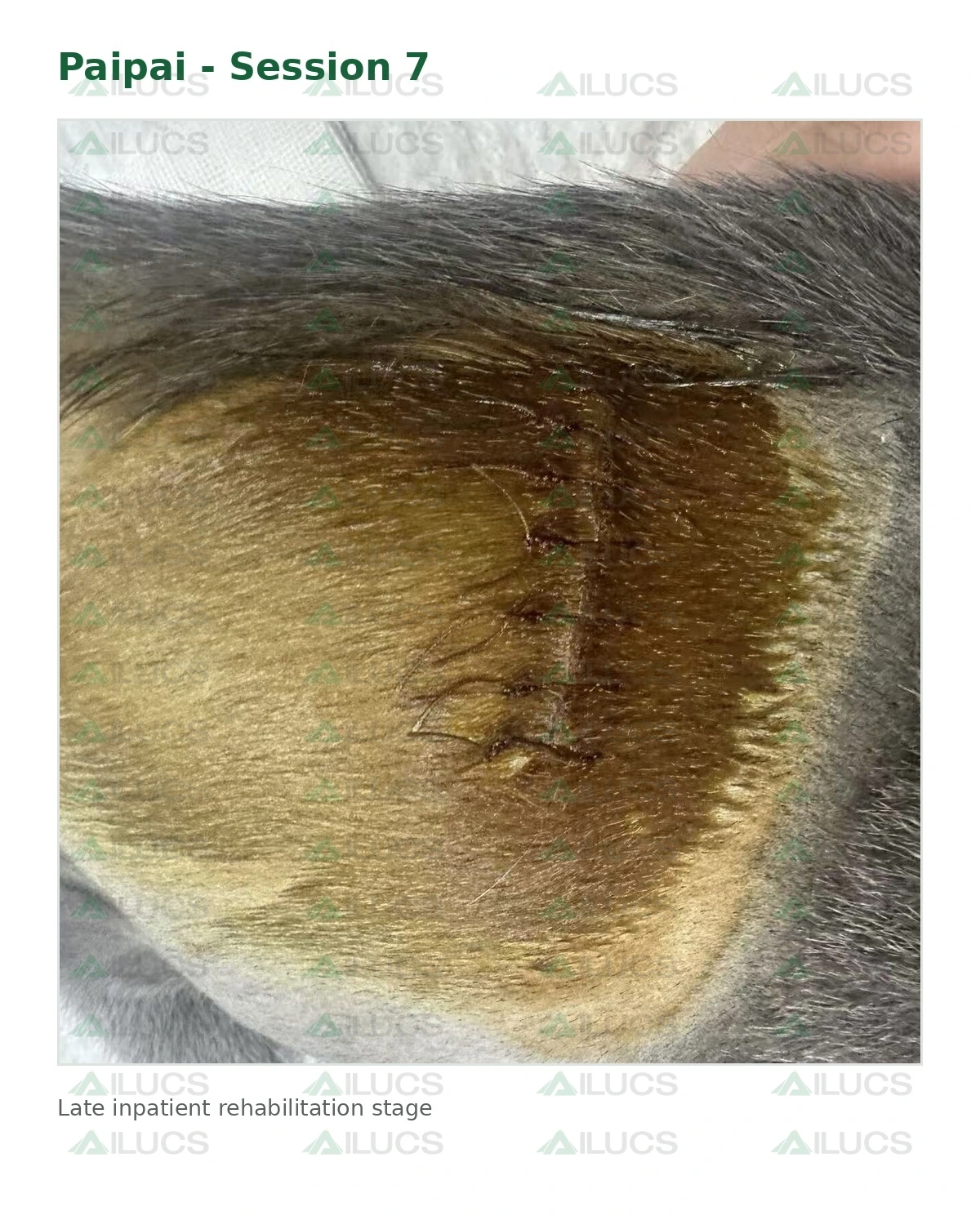
Serial photographs continue the record through Sessions 4-7. Across the later photographs, the sutured incision remains closed and the external appearance of the surgical site changes progressively over time. Because the available material does not include formal wound scores, laboratory data, or a postoperative imaging series, the article should describe only visible changes and documented functional observations rather than assigning numerical healing rates or attributing recovery to laser therapy alone.
6. Functional Recovery and Discharge
The clearest documented functional milestone is Paipai standing independently after the third treatment session, as shown in the early-recovery video. Rehabilitation then continued through seven inpatient laser therapy sessions before discharge.
7. Clinical Interpretation
Paipai’s case illustrates how veterinary laser therapy can be integrated into postoperative orthopedic rehabilitation. Her recovery occurred in the context of surgical treatment, inpatient monitoring, wound care, activity management, time, and repeated clinical reassessment. The observed improvement should therefore not be attributed to laser therapy alone. In this case, laser therapy was used as an adjunctive tool to support local tissue care, comfort and participation in early functional recovery.
8. Takeaway
Paipai’s case documents a right femoral head fracture followed by surgical excision, postoperative laser therapy, and serial observations of incision appearance and early mobility. Independent standing was documented after the third session, and inpatient rehabilitation continued through Session 7 before discharge. These observations illustrate a multimodal postoperative care pathway; they do not establish that laser therapy alone produced the reported recovery.