
Laser Therapy for Reptiles: Safety Considerations for Wound Healing and Tissue Recovery
Laser therapy, also known as photobiomodulation (PBM), is increasingly used in veterinary rehabilitation to support tissue recovery and wound healing. But can the same approach used in dogs and cats be applied to reptiles?
The answer requires careful consideration.
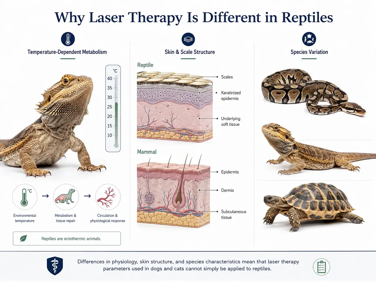
Reptiles are ectothermic animals, meaning their metabolism, immune function, tissue repair, and energy utilization are strongly influenced by environmental temperature. Their scales and skin structure also interact with light differently from mammalian tissue. In addition, reptiles may show subtle or delayed signs of pain and discomfort.
For these reasons, veterinary laser therapy for reptiles should be approached differently from laser therapy in dogs and cats. Treatment parameters should be individualized according to the species, clinical condition, treatment site, and observed response.
This guide explains the key safety considerations for laser therapy in reptiles, including patient preparation, treatment parameters, sensitive anatomical areas, wound care, and long-term monitoring.
Why Is Laser Therapy Different in Reptiles?
Temperature-Dependent Metabolism
Reptile metabolism and tissue repair are closely associated with body temperature. When environmental temperature is inadequate, circulation, oxygenation, and tissue repair may be affected.
Before laser therapy, the patient should be stabilized within its species-appropriate Preferred Optimum Temperature Zone (POTZ). Treatment response should also be interpreted in the context of the animal's current physiological condition.
Maintaining an appropriate thermal environment is therefore an important part of preparing a reptile patient for rehabilitation.
Differences in Skin and Scales
Reptile skin generally has a keratinized epidermis and scales. Compared with mammalian skin, these structures can affect how light is scattered and absorbed.
This means that effective light penetration and delivered energy may differ from what is expected in mammalian patients. Treatment should therefore be considered according to the species, treatment site, and condition rather than relying on a standard mammalian protocol.
Significant Species Variation
“Reptiles” are not a single uniform patient group.
Snakes, lizards, turtles, tortoises, and geckos can differ substantially in skin thickness, scale characteristics, body size, tissue structure, and thermoregulatory requirements.
There is no universal laser therapy protocol suitable for every reptile species. Individual assessment is essential.
Subtle Pain and Behavioral Responses
Reptiles may not display obvious signs of pain or discomfort. A patient that appears calm during treatment may still require careful monitoring.
Veterinarians should therefore assess treatment response using clinical examination, lesion appearance, treatment records, and follow-up observations rather than relying on behavior alone.
Preparing a Reptile Patient for Laser Therapy
1. Confirm an Appropriate Environmental Temperature
Before treatment, confirm that the animal is stable within its species-appropriate POTZ.
Appropriate environmental conditions support normal metabolic activity and tissue repair and can make treatment response easier to evaluate.
If the animal is not within an appropriate thermal range, environmental stabilization should be addressed before interpreting the expected response to laser therapy.
2. Establish a Clear Diagnosis
The condition should be assessed before treatment. A lesion may be a traumatic wound, ulcer, postoperative incision, infectious lesion, or necrotic lesion such as oral ulceration or scale necrosis.
Different conditions require different treatment strategies.
Laser therapy should be considered as part of an overall clinical management plan. It should not replace appropriate wound management, infection control, or treatment of the underlying condition.
3. Assess Potential Contraindications
Areas with suspected or confirmed malignant lesions, radiosensitive gonadal regions, or an acute bleeding tendency require particular caution.
Depending on the clinical situation, direct irradiation may need to be avoided or performed only after appropriate veterinary assessment.
When treatment suitability is uncertain, the underlying condition should be evaluated before laser therapy is initiated.
4. Use Appropriate Restraint and Eye Protection
Reptiles have very different handling requirements. Turtles may retract into their shells, snakes may coil or move unpredictably, and lizards may use their tails defensively.
Appropriate restraint helps minimize movement and reduces the risk of accidental irradiation or secondary injury.
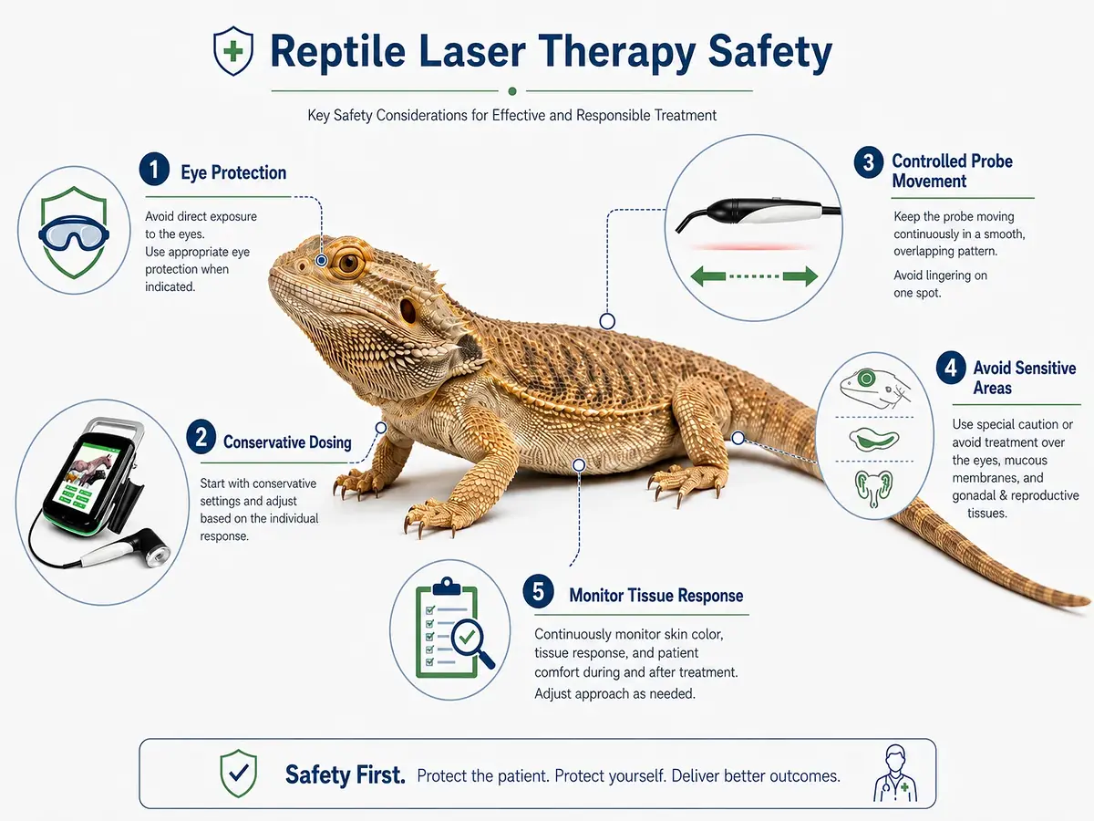
The operator should also wear protective eyewear appropriate for the wavelength being used, particularly when near-infrared wavelengths are applied.
Safe Laser Therapy Practices During Treatment
1. Start Conservatively and Individualize Treatment
Laser therapy for reptiles should begin conservatively rather than assuming that a protocol developed for dogs or cats can be directly applied.
When selecting treatment parameters, consider:
· Species and body size
· Treatment site
· Type and severity of the lesion
· Skin and scale characteristics
· Current physiological condition
· Previous treatment response
For a species with limited clinical experience, starting with a small treatment area and a conservative dose can provide an opportunity to observe the initial response before making further adjustments.
The principle is simple: start conservatively, monitor carefully, and adjust according to the individual patient.
2. Avoid Localized Heat Accumulation
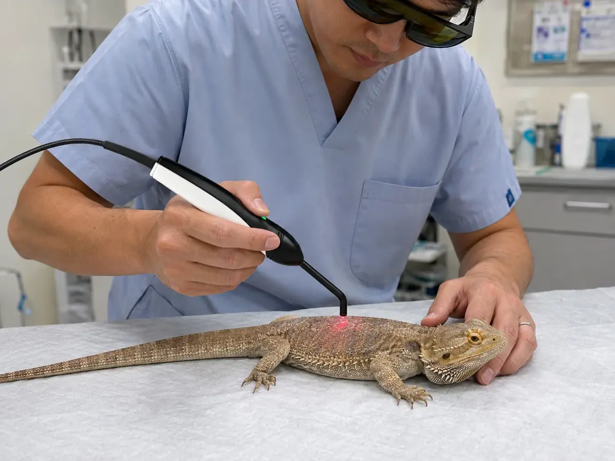
The treatment probe should be moved at a controlled and consistent speed rather than remaining stationary over one location for an extended period.
Although reptiles have different thermoregulatory mechanisms from mammals, localized heat accumulation can still create a risk of thermal injury.
Careful probe movement and appropriate treatment parameters are therefore important parts of safe application.
3. Protect Sensitive Anatomical Areas
Eyes
Protect or avoid the eyes during treatment, particularly in light-sensitive or nocturnal species.
Mucous Membranes and Cornea
These tissues require additional caution. Direct irradiation should be avoided where appropriate or performed using a more conservative approach.
Gonads and Developing Reproductive Tissues
Avoid unnecessary or aggressive irradiation around the gonads and developing reproductive tissues.
4. Monitor the Treatment Area and Patient Response
During treatment, monitor the treatment area for excessive redness, discoloration, blistering, or other unexpected reactions.
If an abnormal reaction occurs, stop treatment and reassess the patient.
Importantly, reptiles may not always show obvious external signs when exposed to excessive energy. The absence of a visible reaction does not necessarily mean that the treatment dose is appropriate.
This is why clinical observation and objective follow-up are important throughout the treatment process.
5. Allow Sufficient Time Between Treatments
Reptile tissue repair may take longer than expected compared with common mammalian patients.
Treatment schedules should therefore be conservative, allowing sufficient time for tissues to respond and recover between sessions.General factors affecting veterinary laser therapy session duration include treatment area, power, dose, and patient response, although reptile treatment schedules still require species-specific assessment.
Photographs and clinical records should be taken before and after treatment whenever possible. Consistent documentation can help evaluate changes in wound size, tissue appearance, ulcer closure, and overall healing progression.
Post-Treatment Care for Reptile Patients
Maintain Appropriate Temperature and Humidity
Appropriate environmental conditions remain important after laser therapy.
Continue to maintain the animal within its species-appropriate temperature range. For species with specific humidity requirements, environmental humidity should also be monitored to support an appropriate healing environment.
Continue Appropriate Wound Care
Laser therapy does not replace standard wound management.Similar principles also apply when integrating veterinary laser therapy into postoperative recovery, where laser treatment should complement wound cleaning, infection control, and routine clinical care.
Depending on the condition, appropriate care may include cleansing, debridement, infection control, and sterile dressing changes.
For open wounds, monitor for signs of infection or deterioration, including increased discharge, abnormal odor, tissue discoloration, or delayed healing.
Laser therapy should be integrated into the overall clinical management plan when appropriate.
Monitor Long-Term Progress
Reptile tissue repair can be relatively slow. A lack of obvious improvement after several treatment sessions should not automatically lead to an aggressive increase in laser dosage.
Instead, treatment response should be evaluated over an appropriate period based on the species, condition, and healing process.
Objective follow-up records are particularly useful when clinical changes are gradual.
For broader information on treatment follow-up and sustained outcomes, see our review of the long-term efficacy of laser therapy in animal rehabilitation.
A Practical Approach for Veterinary Clinics
A structured workflow can help veterinary teams use laser therapy more consistently when treating reptile patients.
Before Treatment
Confirm:
· Species and relevant husbandry requirements
· Stable environmental temperature
· Diagnosis and treatment objective
· Condition of the treatment site
· Sensitive or contraindicated areas
During Treatment
During treatment, record the main laser treatment parameters and clinical observations, including wavelength, power, treatment mode, treatment duration, treatment area, and patient response.
These records can help veterinarians compare treatment sessions and make future adjustments more systematically.
During Follow-Up
Use standardized photographs and clinical notes whenever possible.
Rather than relying only on whether a wound “looks better,” compare objective changes over time, such as wound size, tissue appearance, ulcer closure, and overall healing progression.
This is particularly useful for reptile patients because recovery may be gradual and visible changes can be subtle.
Reptile Laser Therapy Safety Checklist
| Treatment Stage | Key Considerations |
| Before treatment | Stabilize temperature, establish a diagnosis, and assess contraindications |
| Treatment parameters | Start conservatively and individualize according to species and condition |
| Wavelength / probe | Use appropriate parameters, maintain controlled probe movement, and avoid heat accumulation |
| Sensitive areas | Protect or avoid the eyes, mucous membranes, cornea, gonads, and developing reproductive tissues |
| Animal handling | Use appropriate restraint and minimize unnecessary movement |
| Operator safety | Wear wavelength-specific protective eyewear |
| During treatment | Monitor skin and patient responses and stop if abnormal reactions occur |
| After treatment | Maintain appropriate temperature and humidity, continue wound care, and monitor healing |
Frequently Asked Questions About Laser Therapy for Reptiles
1.Can laser therapy be used on reptiles?
Laser therapy may be considered as part of a veterinary rehabilitation or wound management plan for reptile patients. However, treatment should be individualized according to the species, clinical condition, treatment site, and environmental conditions.
2.Can I use the same laser parameters for reptiles and dogs or cats?
Treatment parameters should not simply be transferred from dogs or cats to reptiles. Differences in metabolism, skin structure, thermoregulation, and species-specific responses should be considered when developing a treatment approach.
3.What should be checked before reptile laser therapy?
The patient's environmental temperature, diagnosis, treatment site, potential contraindications, and restraint requirements should be assessed before treatment.
4.What should I monitor during treatment?
Monitor the treatment area for abnormal redness, discoloration, blistering, or other unexpected reactions. Patient behavior and overall response should also be observed.
5.Does laser therapy replace wound care?
No. Laser therapy does not replace appropriate wound management, infection control, or treatment of the underlying condition. It should be used as part of an appropriate clinical management plan.
6.How quickly should reptile wounds respond to laser therapy?
Healing can be gradual in reptiles. A lack of immediate visible improvement should not automatically lead to a rapid increase in treatment intensity. Treatment response should be evaluated over an appropriate period using objective clinical records.
Final Thoughts: Safety Comes First
Reptile patients require a different approach to laser therapy because their physiology, thermoregulation, skin structure, and behavioral responses differ from those of mammals.
The goal should not be to simply adapt a canine or feline laser protocol to a reptile patient. Instead, veterinarians should consider the species, environmental conditions, treatment site, clinical condition, treatment parameters, and individual response when developing a treatment strategy.
Laser therapy can provide a non-invasive and gentle therapeutic option to support wound healing and tissue recovery in reptiles when used appropriately and as part of an overall clinical management plan.
Safe treatment begins with a few basic principles:
· Maintain an appropriate environmental temperature
· Start conservatively and individualize treatment
· Protect sensitive anatomical areas
· Follow appropriate laser and animal-handling safety procedures
· Continue proper wound care
· Monitor outcomes objectively over time
Understanding how reptiles differ from mammals is the first step toward safer and more appropriate veterinary laser therapy.
References
1.Mitchell, M. A., & Diaz-Figueroa, O. (2004). Wound management in reptiles. Veterinary Clinics of North America: Exotic Animal Practice, 7(1), 123–140.
2.Mader, D. R., & Divers, S. J. Principles of wound management and wound healing in exotic pets.
3.Millis, D. L., & Bergh, A. (2023). A systematic literature review of complementary and alternative veterinary medicine: Laser therapy. Animals, 13(4), 667.
4.A systematic review via text mining approaches of human and veterinary applications of photobiomodulation: Focus on multiwave locked system laser therapy. (2025). Lasers in Medical Science, 40, 321.
About AILUCS
AILUCS focuses on veterinary rehabilitation and therapeutic equipment, providing veterinary professionals with laser therapy solutions and multi-species rehabilitation technologies.
Our goal is to support veterinary teams with practical tools that can be integrated into clinical workflows across different animal species.
For reptile patients, as with every species, appropriate treatment begins with understanding the animal and putting safety first.